
Key Takeaways
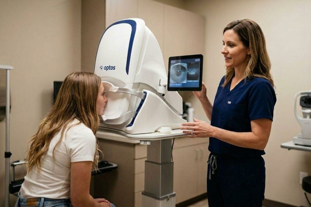
- Optomap captures a wide panoramic view of your retina, showing more than 80% of it in a single image and making it a valuable tool for annual eye exams.
- Optical coherence tomography (OCT) provides a detailed cross-section of retinal layers and is used to detect, diagnose, and monitor certain eye conditions.
- A free OCT scan offered elsewhere does not provide the same wide view of your retina as Optomap imaging or replace a comprehensive eye exam.
- Both tools serve different purposes, and your optometrist may recommend one or both depending on your needs.
Optomap & OCT Are Different
You’ve probably seen clinics advertising free OCT and wondered if it’s a good screening tool or if it’s the same screening technology that you get at an eye exam from our team at Eyes on Weston. It’s a fair question, and the answer matters more than most people realize.
Optomap and OCT are two separate diagnostic tools that do different jobs. Knowing the difference can help you make more informed decisions about your family’s eye care.
Two Different Tools, Two Different Jobs
Optomap: A Wide-Angle View of Your Retina
Optomap captures a wide, panoramic image of your retina, allowing us to view more than 80% of it in a single image, including areas toward the outer edges (periphery) of your retina. At Eyes on Weston, your Optomap imaging also includes an autofluorescence image, which provides additional information about the health of your retina including inflammatory disease, hereditary retinal disorders and early signs of age-related macular degeneration.
OCT: A Detailed Look at Your Macula
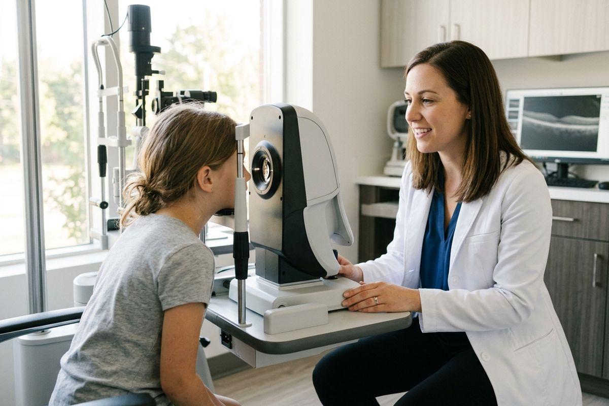
OCT (optical coherence tomography) takes a detailed, cross-sectional image of the different layers of your macula, the central part of your retina responsible for sharp, detailed vision. These scans allow us to examine the retinal layers closely and identify subtle changes of retinal disease. However, OCT only provides information about the specific areas scanned and does not give insight into the peripheral retina.
Why Are Both Important?
Optomap and OCT complement each other by providing different views of your retinal health. Optomap gives us a broader view of your retina, while OCT allows us to examine specific structures in greater detail.
Optomap is a more comprehensive screening tool for the overall retina because it captures a wide, panoramic view of the retina, including the peripheral areas where many retinal conditions may develop. In contrast, a macular OCT examines detailed cross-sections of specific central retinal structures better used to monitor pre-existing conditions.
Using both technologies helps us gain a more complete picture of your eye health, because no single imaging test provides all the information needed to assess every part of your retina.
What Optomap Shows at Your Annual Eye Exam
A Wide View for Annual Checkups
Optomap gives your optometrist a broad look at your retina in one comfortable, quick image. In most cases, no dilation drops are needed, which makes the process easy for both children and adults. Because Optomap covers so much of the retina at once, it’s well-suited for routine annual eye exams where the goal is to catch changes you may not feel or notice yet.
Optomap images can also be saved and compared over time, helping your optometrist identify changes in your retinal health. However, dilation is still recommended.
Conditions Optomap Can Help Identify
During a routine annual exam, Optomap can help your optometrist spot signs of a range of different conditions. These include:
- Diabetic retinopathy and retinal bleeds in people with diabetes.
- Changes in retinal blood vessels that may be associated with conditions such as high blood pressure.
- Retinal tears and detachments, which may be associated with symptoms such as floaters and flashing lights.
- Retinal freckles and other lesions that need monitoring.
- Hereditary retinal disorders that occur during development, such as optic nerve head drusen, retinitis pigmentosa, and stargardt disease.
- Macular retinal disease such as age-related macular degeneration, and medication toxicity.
What OCT Shows & When It’s Recommended
A Closer Look at Retinal Layers
Where Optomap gives breadth, OCT gives detail. OCT maps the individual layers of your retina in precise structural detail, which is why your optometrist may recommend it to detect, diagnose, or monitor certain eye conditions. Glaucoma, age-related macular degeneration (AMD), and fluid buildup in the retina are among the conditions that OCT can help identify and monitor over time. At Eyes on Weston, our screening Optomap includes a series of OCT scans as part of our screening process, however a more detailed scan may be needed to monitor change and progression of certain eye conditions.
OCT is particularly useful for examining specific retinal structures in detail. However, a typical OCT scan doesn’t capture the same broad view of the peripheral retina that Optomap provides.s.
Free OCT: What It Does and Does Not Cover
When a clinic advertises free OCT, this can sound like a thorough eye health checkup. In practice, however, a typical OCT scan focuses on a specific area of your retina rather than capturing the broad panoramic view that Optomap provides. Because some retinal abnormalities develop outside the central retina, ultra-widefield imaging can offer valuable additional information during routine eye exams.
Free OCT can provide useful diagnostic information, but it isn’t an effective screening tool. When comparing eye care options, it’s worth asking what retinal imaging is included and how your optometrist assesses your overall eye health.
Why Vaughan Families Benefit from Optomap at Annual Exams
One of the practical strengths of Optomap is how well it works across age groups. It’s fast and comfortable, which matters when you’re bringing children in for their annual appointments. For families with a history of eye disease, myopia, or diabetes, that broad annual retinal view can help identify and monitor changes that may develop before noticeable symptoms appear.
Its ability to capture images of both the central and peripheral retina also gives your optometrist a broader picture of your retinal health, helping support personalized care and ongoing monitoring. At Eyes on Weston, we start Optomap screening at the age of 10, unless advised earlier.
OCT & Optomap Together
Your optometrist may recommend both Optomap and OCT together for:
- Retinal abnormalities that require a closer examination of specific structures.
- Tracking how a diagnosed eye disease is responding to treatment.
- Cases where combined imaging provides complementary information about your retinal health.
Choosing the Right Eye Doctor in Vaughan for Modern Eye Care
Personalized care means the right imaging for your situation, whether that’s Optomap, OCT, or both. At Eyes on Weston by Gagliardi Optometric Clinic, that kind of thoughtful, individualized approach is what families across Vaughan and the York region have come to count on.
With its ability to capture more than 80% of the retina in a single image, Optomap offers a valuable advantage for broad retinal screening during routine eye exams.
To learn more or to book your family’s next eye exam, reach out to our team at Eyes on Weston today.